Pelvic Anatomy - Female Pelvic Anatomy Artwork Photograph By Science Photo Library - Welcome to the valuemd albums.. The pelvis (plural pelves or pelvises) is either the lower part of the trunk of the human body between the abdomen and the thighs (sometimes also called pelvic region of the trunk) or the skeleton embedded in it (sometimes also called bony pelvis, or pelvic skeleton). 3d interactive models and tutorials on the anatomy of the abdomen and pelvis. The pelvic girdle consists of two symmetrical halves. Interactive video showing normal female pelvic anatomy as seen by laparoscopy. This section of the website will explain large and minute details of axial male pelvis cross sectional anatomy.
Register now and grab your free ultimate anatomy study guide! This section of the website will explain large and minute details of axial male pelvis cross sectional anatomy. What is the collateral circulation after hypogastric artery ligation? We'll go over the main differences and dive into the anatomy and function of the different parts of the female uterus. The female pelvis is slightly different from the male pelvis.
3d Rendered Medically Accurate Illustration Of The Pelvic Anatomy Stock Photo Picture And Royalty Free Image Image 138357661 from previews.123rf.com Anatomy pelvis muscles pubococcygeus, puborectalis and iliococcygeus., pelvis nerve, the spinal nerves that arise from. The pelvic girdle consists of two symmetrical halves. I've been putting this together for a while and am very excited to share this with you! 3d interactive models and tutorials on the anatomy of the abdomen and pelvis. Introduction the anatomical basis of pelvic floor function in normal and abnormal states pelvic floor ultrasound: Branches of the internal iliac artery. Learn about the blood vessels, organs, nerves and peritoneal cavity. The female pelvis is slightly different from the male pelvis.
Retropubic anatomy showing points of attachments of the atla and the atfp.
Surgical pelvic anatomy in gynecologic oncology. Interactive video showing normal female pelvic anatomy as seen by laparoscopy. Pelvic skeleton includes two hip bones, sacrum and coccyx. Introduction the anatomical basis of pelvic floor function in normal and abnormal states pelvic floor ultrasound: Laparoscopic understanding of pelvic anatomy and its application in benign and radical pelvic surgery. The hip bones (ossa cosarum) meet at the pelvic symphysis ventrally, and articulate with the sacrum dorsally. Laparoscopic anatomy of the female pelvic region. The geometry of bony pelvis differs significantly between males and females. Journal of anatomy and physiology, vol. Agreements & disagreements workshop 36. This mri pelvis cross sectional anatomy tool is absolutely free to use. Pelvic floor anatomy in 3d (as it should be). The female pelvis is slightly different from the male pelvis.
Choose from 500 different sets of flashcards about pelvic anatomy on quizlet. Provides excellent detail of bony anatomy and can confirm pelvic ring / acetabular fractures that are not always visible on plain radigraphs. Pelvic skeleton includes two hip bones, sacrum and coccyx. Anatomy of pelvis & perineum by profgoodnewszion 71948 views. Agreements & disagreements workshop 36.
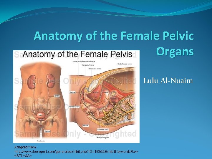
Anatomy Of The Female Pelvic Organs Lulu Alnuaim from slidetodoc.com Ever wanted a resource with articles, pictures, and videos of pelvic anatomy all int he same place? There are many organs that sit in the pelvis, including much of the urinary system, and lots of the male or female reproductive systems. Bony pelvis the bony pelvis is formed by the sacrum and coccyx and a pair of hip bones (ossa android pelvis (20%): Pelvic floor anatomy & function: What is the collateral circulation after hypogastric artery ligation? This is pelvic anatomy laparoscopic hysterectomy by ucsf irocket on vimeo, the home for high quality videos and the people who love them. The lesser or true pelvis (pelvis minor).—the lesser pelvis is that part of the pelvic cavity which is situated below and behind the pelvic note 59. Learn about pelvic anatomy with free interactive flashcards.
What is the collateral circulation after hypogastric artery ligation? Pelvic floor anatomy & function: Welcome to the valuemd albums. The bony pelvis & gender differences in pelvic anatomy. Agreements & disagreements workshop 36.
Clinical Pelvic Anatomy Clinical Obstetrics And Gynaecology 3ed from doctorlib.info Provides excellent detail of bony anatomy and can confirm pelvic ring / acetabular fractures that are not always visible on plain radigraphs. Interactive video showing normal female pelvic anatomy as seen by laparoscopy. Pelvic floor anatomy & function: The measurements of each of these regions is important as the fetal head has to negotiate its way through. The pelvis (plural pelves or pelvises) is either the lower part of the trunk of the human body between the abdomen and the thighs (sometimes also called pelvic region of the trunk) or the skeleton embedded in it (sometimes also called bony pelvis, or pelvic skeleton). The true pelvis is divided into three regions known as the pelvic brim, the cavity and the outlet. Register now and grab your free ultimate anatomy study guide! Learn about pelvic anatomy with free interactive flashcards.
Pelvic floor anatomy & function:
We'll go over the main differences and dive into the anatomy and function of the different parts of the female uterus. Laparoscopic anatomy of the female pelvic region. Female pelvis ppt by mayil rasamani 144734 views. This mri pelvis cross sectional anatomy tool is absolutely free to use. Introduction the anatomical basis of pelvic floor function in normal and abnormal states pelvic floor ultrasound: Bony pelvis the bony pelvis is formed by the sacrum and coccyx and a pair of hip bones (ossa android pelvis (20%): Pelvic floor anatomy in 3d (as it should be). The true pelvis is divided into three regions known as the pelvic brim, the cavity and the outlet. The measurements of each of these regions is important as the fetal head has to negotiate its way through. Ever wanted a resource with articles, pictures, and videos of pelvic anatomy all int he same place? Welcome to the valuemd albums. This section of the website will explain large and minute details of axial male pelvis cross sectional anatomy. Branches of the internal iliac artery.


0 Comments:
Post a Comment